DOE Hyperspectral Imaging for Cancer Detection
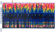 |
This technology using noninvasive hyperspectral imaging may detect cancerous and precancerous abnormalities in human tissue. The 3-dimensional optical imaging system employs high spectral resolution and narrow bandwidths to create highly detailed images that distinguish cancerous tissue from healthy tissues. A comparison of these tissue segments and cells permits diagnosis and delineation of suspect tissue.
Hyperspectral imaging can be used to detect cancerous tissues in all areas of the human body accessible through endoscopy:
- Oral cavity cancers – tongue, gum, mouth, tonsil, salivary gland
- Digestive system cancers – colorectal, stomach, esophagus, small intestine
- Respiratory system cancers – lung, bronchus, pleura, larynx
- Female genital cancers – corpus, ovary, vulva, uterus
This highly detailed 3-D imaging measures colors simultaneously in hundreds of spectral bands, instead of just a handful of spectral bands as with current technology. It provides objective, quantifiable data about the spectral signatures of suspect tissue to reduce reliance on subjective judgments. Other benefits of this technology include:
- Precise imaging reveals details not visible to the human eye or to other imaging techniques
- Image may accurately detect cancerous and precancerous tissue, and further discriminates tissue into multiple tissue types of categories
- Image covers large areas, minimizing risk of overlooking suspect tissue and precisely delineating tumor margins
- Miniature probe size permits noninvasive diagnosis wherever endoscope can be used
“Hyperspectral imaging methods and apparatus for non-invasive diagnosis of tissue for cancer” is covered by U.S. Patent No. 5,782,770 (Dr. Mark Wiederhold, co-inventor), issued July 21, 1998; the automated hyperspectral imaging software is U.S. Patent Pending.
